Virtual MRI
A realistic simulation of a MRI was developed. For the user it should be possible to change all relevant setting of the virtual scanner and to adapt them to the expected pathology. Students and doctors in training are the target group.
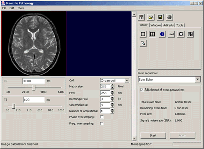
At the moment the pulse sequence classes SR, IR, SE, TSE, FLASH and FISP are implemented. By a plugin mechanism this list can be easily extended. Parameters, like TR, TE, TI, flip-angle or echo train length, can be adjusted. The choice of matrix size, FOV, slice-thickness and number of acquisitions affect the signal-to-noise ratio of the images. In a first step, the simulation calculates the signal intensity in the k-space. Aliasing- and motion-artifacts are simulated by modifying the k-space data. In a last step, a 2D-fourier-transform of the k-space data is performed. Window and center of the resulting images can be changed. The simulation time can be optionally delayed till the real measuring time. The algorithms of the simulation are based on parameter-images of the three physical basic items T1, T2 and proton density. They are calculated once from a patient examination with suitable pulse sequences. To work out pathologies a small graphic tool is included. The graphical user interface is oriented on a real MR scanner. The software is implemented in pure Java under the GPL license.
On a 500 MHz PC the software calculates an image in 5 to 20 seconds, depending on the pulse sequence and the degree of the desired artifacts.
An interactive simulation of a real world examination is possible on a standard PC. The users can study the operation of a costly and not everywhere available equipment on their desktop.
RSNA 2002
The current version of the simulation was presented at the RSNA 2002, 88th Scientific Assembly and Annual Meeting, Chicago, IL, USA. During the meeting a lecture was given. The presentation was given the RSNA infoRAD award cum laude.
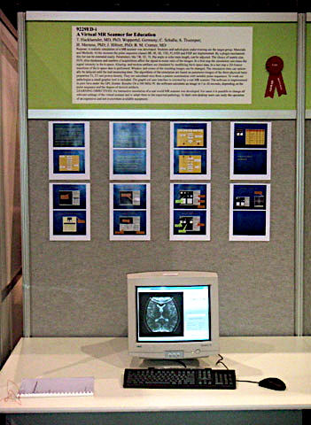
Picture of the booth at the RSNA 2002 infoRAD
Hardware and software requirements
The virtual magnetic resonance scanner is programmed in pure Java. Therefore, the program runs on every computer a Java virtual machine or Java Runtime Environment (JRE), version 1.3 or higher, is installed on.
To perform the extensive calculations of the simulation within an acceptable time a Pentium II processor with a clock speed of 400 MHz and a random access memory (RAM) of 128 MB has to be available under Windows and Linux i386 at least. The system excellently runs on a Pentium III processor with 500 MHz and 256 MB. For a better performance a large amount of RAM is more important than a faster processor.
For the hard disk space 1 MB is sufficient.
If you want directly to start the program from a CD the CD-ROM drive has to be able to read the CD with speed 20X at least.
User Manual
Download the PDF formatted user manual here. Please follow the link to "Virtual MR scanner" .
Software
The software is developed under the GNU General Public License version 2.0 (GPLv2) and can be downloaded here.
Tools
We have developed two additional tools, which are based on ImageJ:
One of these plugins can be used to calculate the parameter images of the Virtual MR from clinical examinations. The other one allows the user to edit pathologies on the parameter images.
Please note that this software is in alpha stage and currently no documentation is available.
You can download the software here. Please follow the links to "Parameter image calculator" or "Pathology editor" .
Project Team
Concept:
Thomas Hacklaender, MD, PhD
Programming:
Christian Schalla
Andreas Truemper
Thomas Hacklaender, MD, PhD
Localization:
English version, Heinrich Mertens, PhD
Estonian version, Katri Hackländer, MD
Documentation:
English version, Heinrich Mertens, PhD
License
The software is distributed under the GPL license.
References
Schalla C, Trümper A: Entwicklung eines virtuellen klinischen Kernspintomographen. Diplomarbeit 1999. Interner Bericht der Universität Dortmund, Fachbereich Informatik.
Hackländer T, Schalla C, Trümper A, Mertens H, Hiltner J: Virtueller Kernspintomograph. 81. Deutscher Röntgenkongreß, Wiesbaden. RoFo Fortschr Geb Röntgenstr Neuen Bildgeb Verfahr 172 (2000) S135.
Hacklaender T, Schalla C, Truemper A, Mertens H, Hiltner J, Cramer BM: A Virtual MR Scanner for Education. RSNA 2002, 88th Scientific Assembly and Annual Meeting, Chicago, USA. Radiology 225 (P) (2002) 757.
Hackländer T, Mertens H, Cramer BM: Computersimulation eines klinischen Magnetresonanztomographen für Ausbildungszwecke. RoFo Fortschr Geb Röntgenstr Neuen Bildgeb Verfahr 176 (2004) 1151-1156
Hackländer T, Mertens H: Virtual MRI: A PC based simulation of a clinical MR scanner. Acad Radiol 12,1 (2005) 85-96